Yeast Images
S. cerevisiae images available from The Cell: An Image Library
![]()
The Cell: An Image Library™ is a public repository of images and videos. These images have been reviewed and many have been annotated. You will find that The Cell provides a large collection of yeast images and videos. The Cell is funded by NIGMS Grand Opportunities grant RC2GM092708 to the American Society for Cell Biology (ASCB). We encourage you to submit images from your work to The Cell.
The following images were previously provided by the yeast community for distribution bu SGD. You are welcome to use these images, however please be respectful and cite the person(s) who provided the image as the source. If you have images you would be willing to make available for public use, please contribute them to The Cell Library mentioned above.
| Saccharomyces cerevisiae images provided by Diane Nowicki and Ryan Liermann | |
| DY150 cells in stationary phase. [electron micrograph] | 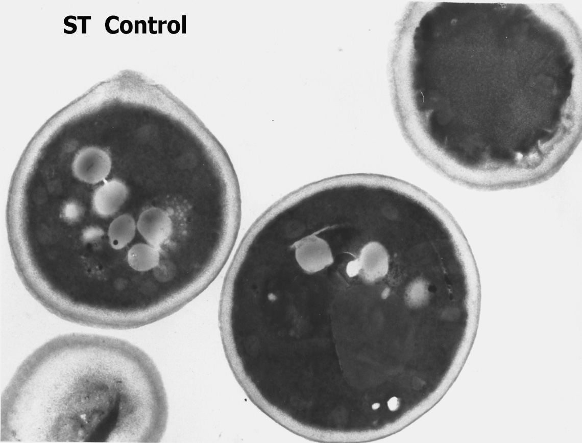
|
| DY150 cells undergoing detergent mediated (Y-PER) lysis. [electron micrograph, 12,000X] | 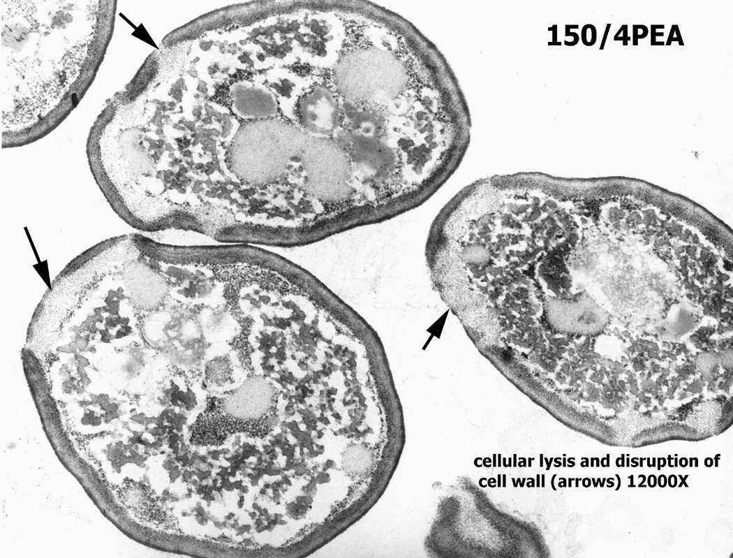
|
| Saccharomyces cerevisiae images provided by Peter Hollenhorst and Catherine Fox | |
| Wild type cells (W303 derivative). [light micrograph] | 
|
| fkh1 delete cells (W303 derivative). [light micrograph] | 
|
| fkh2 delete cells (W303 derivative). [light micrograph] | 
|
| fkh1 fkh2 double delete cells showing pseudohyphal growth (W303 derivative). [light micrograph] | 
|
| Saccharomyces cerevisiae images provided by Carsten Kettner | |
| Wild type YCC588 cells stained with Calcofluor White. [fluorescent micrograph, 1000X] | 
|
| Wild type YCC588 protoplasts. [DIC image, 400X] |  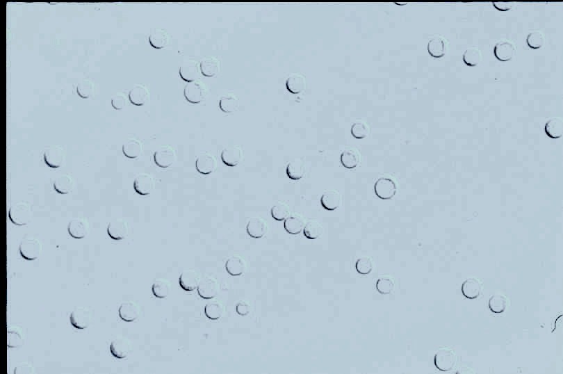
|
| Wild type YCC588 protoplasts. [DIC image, 400X] | 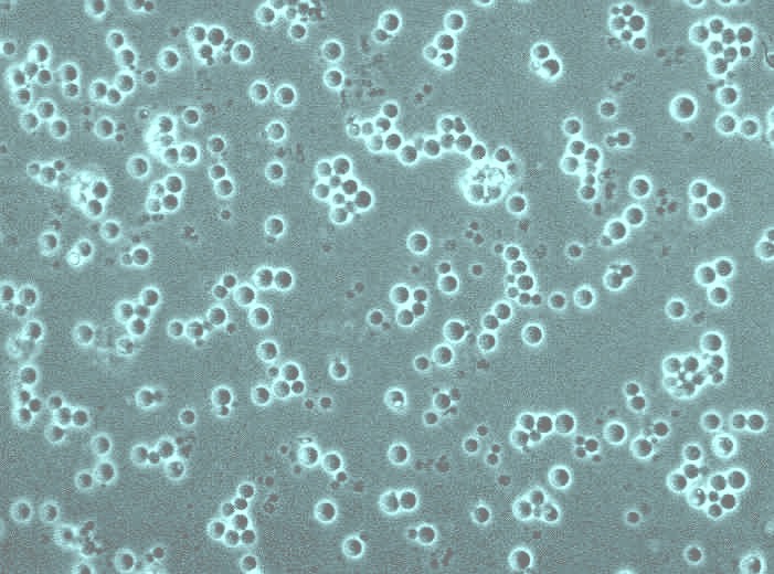
|
| Saccharomyces cerevisiae images provided by Dan Eshel | |
| Axial-budding cells with oval, rounded mother tips. | 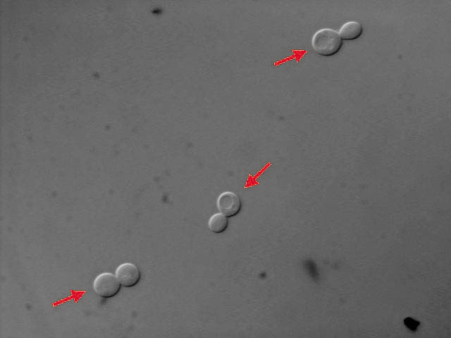
|
| Bi-polar budding cells with "lemon"-shaped mother tips. | 
|
| Saccharomyces cerevisiae image provided by Teresa Rinaldi | |
| Wild type W303 cells, with GFP imported into mitochondria and DNA stained with dapi. [fluorescence microscopy image, 100X] | 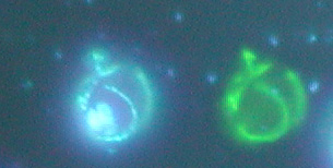
|
| Saccharomyces cerevisiae movies provided by Kerry Bloom | |
| Fluorescent tufts of kinetochore microtubules shortening to the pole. | 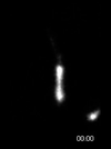
|
| gRNA-ASH1 in bud6 cells. (Strain JZY1345) | |
| Kar9-GFP in WT (Strain YEF473A). | 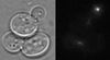
|
| Bloom Lab | |
| Saccharomyces cerevisiae movie provided by Michel Jacquet | |
| Oscillatory nucleocytoplasmic shuttling of Msn2-GFP in W303. | 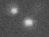
|
| Schizosaccharomyces pombe pictures available from the Forsburg lab page | |
| Saccharomyces cerevisiae images provided by Maxim Zakhartsev | |
| Wild type Saccharomyces cerevisiae strain BY4742 (EUROSCARF) in continuous culture at 22.5 degrees C. Instrument: confocal laser microscope Carl Zeiss LSM 510 Meta, phase contrast imaging. Authors: Dr. Maxim Zakhartsev and Doris Petroi, International University Bremen, Germany. | 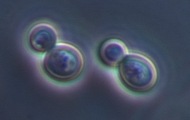
|
| Saccharomyces cerevisiae strain InvSC1 (Invitrogen) transformed with pYES2-eGFP expressing intracellularly recombinant eGFP (enhanced green fluorescent protein from jellyfish Aequorea victoria). Top, large eGFP fluorescence image; Bottom, composite image: upper left, eGFP fluorescence; upper right, phase contrast image; lower left, superimposed image. Instrument: confocal laser microscope Carl Zeiss LSM 510; Meta. eGFP has been excited by argon laser at 488 nm. Authors: Dr. Maxim Zakhartsev, Dr. Sergei Rarozin, Carmen Momeu, International University Bremen, Germany. | 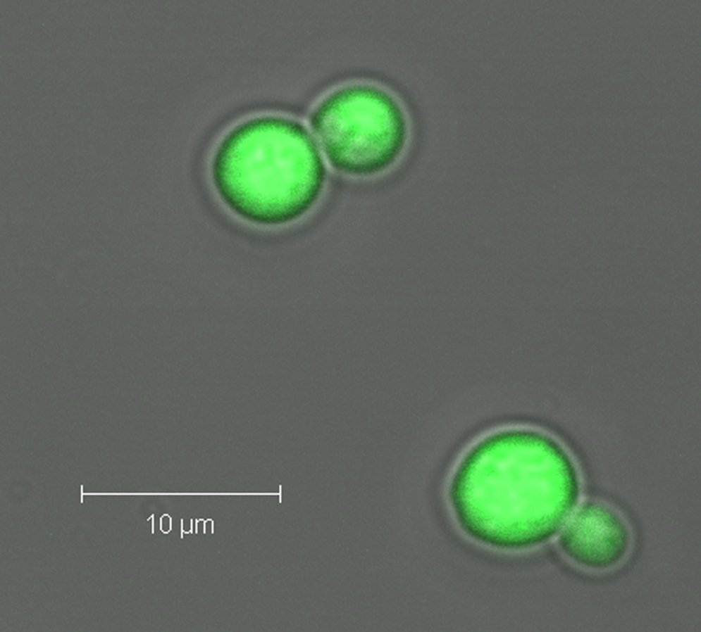
|