UW-Stout/Bud Scars FA21
Measuring The Number of Yeast Cell Bud Scars Through Calcofluor White Staining
Introduction
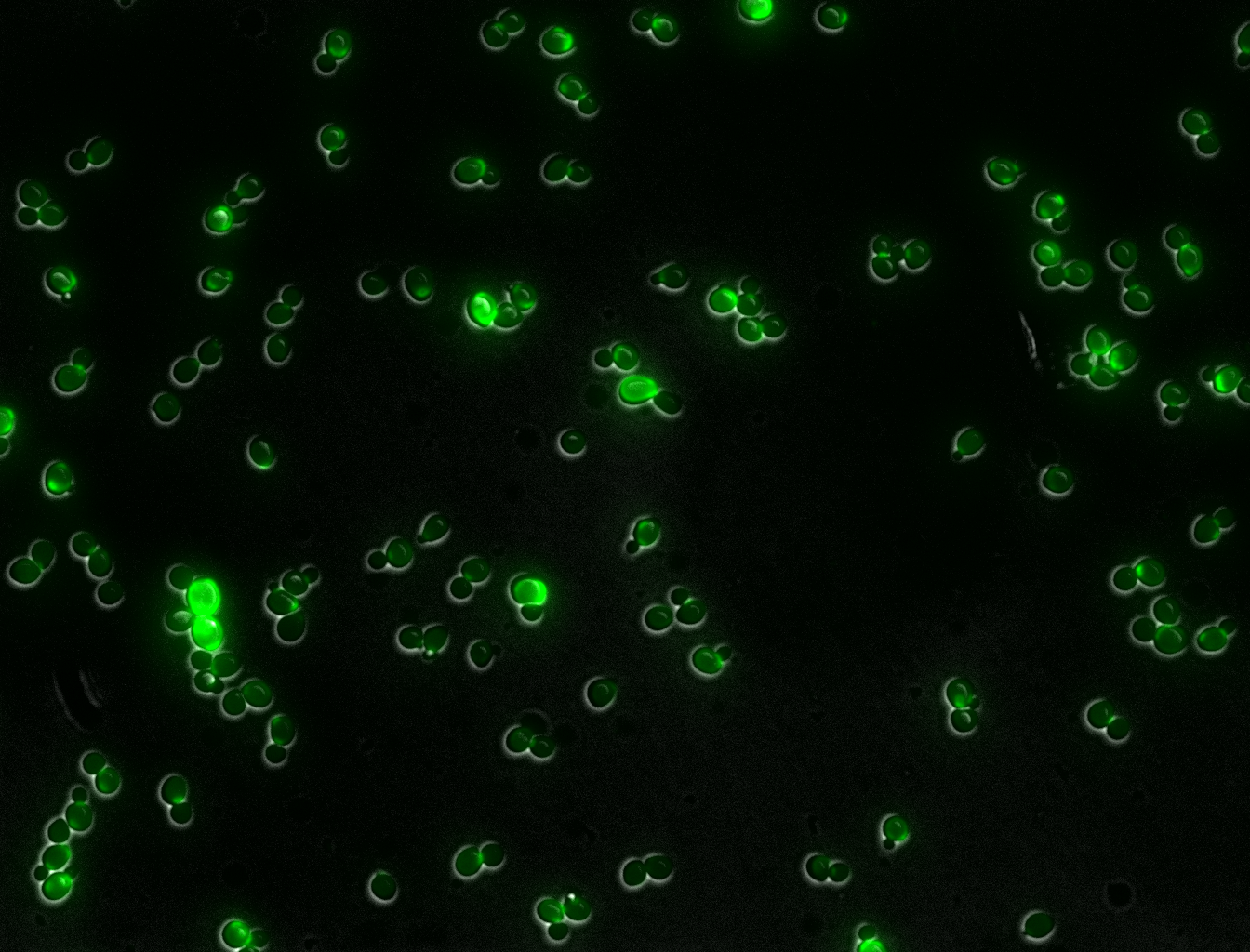
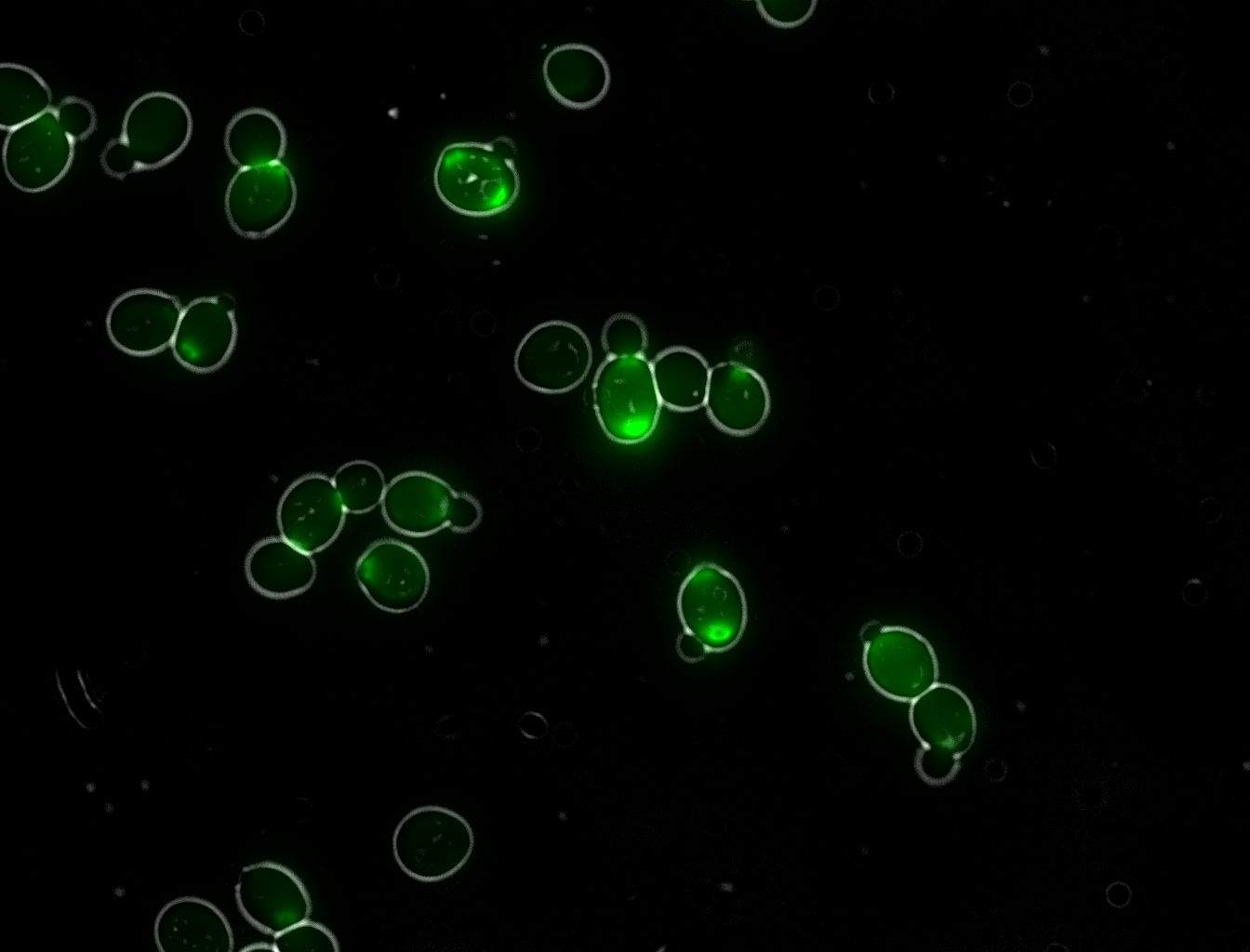
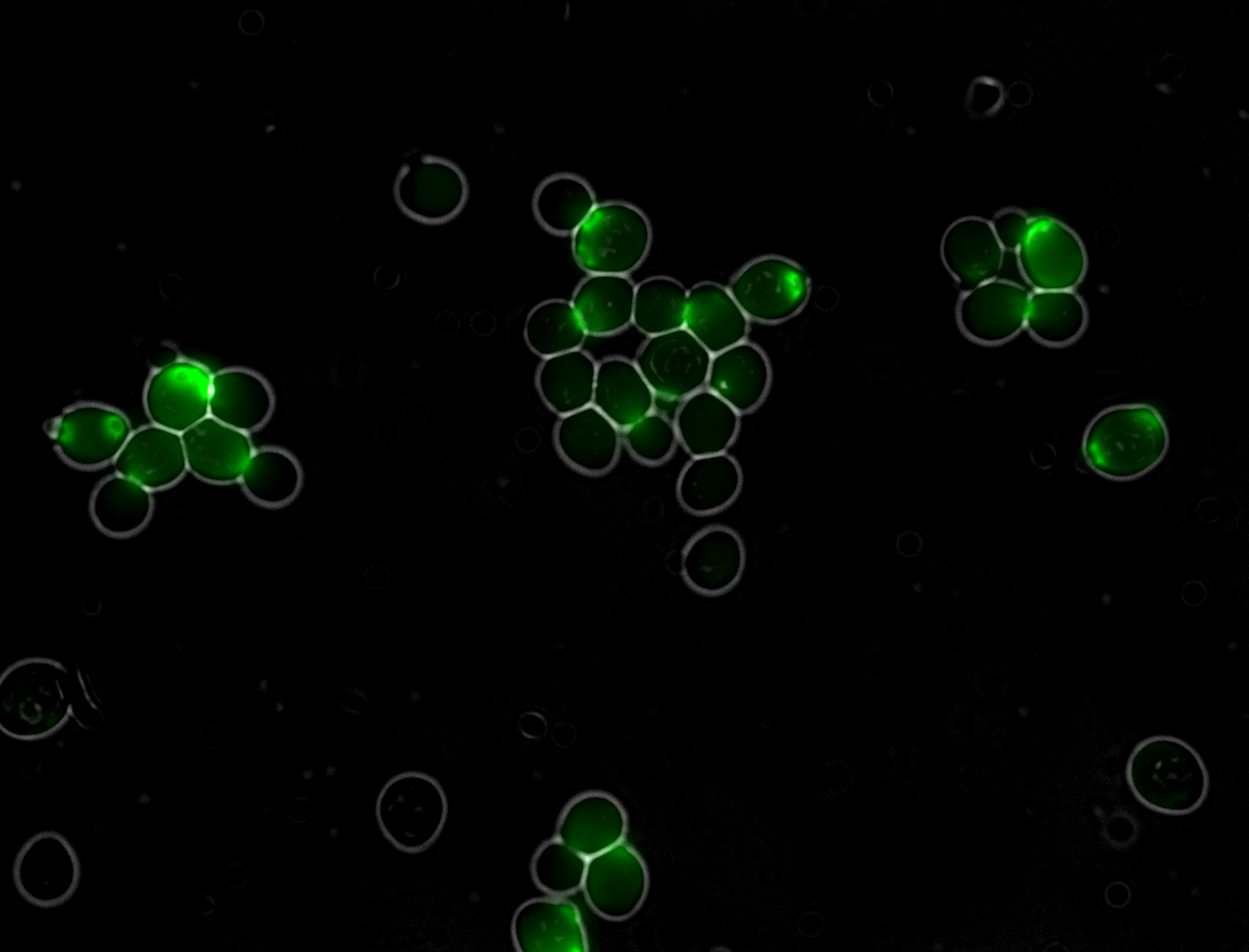
When a yeast cell produces a bud that goes on to become another viable yeast cell, the cleavage of the daughter cell from the parent during cytokinesis leaves a bud scar. These bud scars can be viewed using fluorescent microscope techniques. The number of bud scars visible on each cell can be counted as an indication of how many offspring the cell has had.
The present experiment is designed to allow wild type (WT) and the gene knockout yeast culture to naturally reproduce and then stain these cells with the calcofluor white stain kit so that the number of scars present in the cells can be estimated for each colony. The cultures will be viewed under a microscope and the average number of bud scars per 100 cells will be quantified. Three separate researchers will count the number of scars visible and average these numbers to quantify the number of offspring each yeast cell has. The hypothesis is that there will be a measurable difference between WT and knockout gene yeast cells.
Materials
WT Yeast Culture
Gene Knockout Yeast Culture
1.7ml microcentrifuge tube
Calcofluor White Stain Kit Reagent B- Calcofluor White (1.0 g), Evans Blue Dye (0.4 g), and Demineralized Water (1000.0 ml)
Glass Microscope Slide
Coverslip
Microscope
Procedure
1. Acquire all necessary materials, i.e., remove transformed yeast cells from incubator, etc.
2. Fix the cells by adding formaldehyde 3.7% (115.6ul) to 1ml of yeast cells. Vortex briefly and let this sit for 45-60 minutes.
3. Centrifuge this tube to make a pellet, then wash with water (1ml) twice. The washing procedure is below.
-Centrifuge at 13k for 60s
-Add 1ml water, vortex, centrifuge, remove liquid without disrupting pellet, repeat once more
4. Pellet this and suspend in 10ul of calcofluor.
5. Incubate this for 5 min at room temperature.
6. Pellet cells (centrifuge) and wash with water again following the above procedure.
7. resuspend the final pellet in 50ul of water.
8. Mount on slide and cover with coverslip.
-Place petroleum jelly around the edges of the coverslip to prevent moisture escape.
9. Put a paper towel below and above and cover with a heavy object (like a book) for 10 minutes.
10. Look at the cells under the microscope and observe data.
-For each sample, view 100 different cells and count the number of bud scars.
-For this experiment, 3 researchers simultaneously viewed the microscope image on a screen. An area of cells would be viewed, and each researcher would count the number of cells and number of bud scars seen. If the numbers matched, then they would be recorded, and a new area of the slide would be viewed, and the counting procedure repeated.


